Monthly Archives: May 2017

Is the X-ray diffraction theory we use correct?

The calculated intensities as Ω and X are varied for a detector at 2θ = 60° (a) and 2θ = 110° (b) for 10 µm crystallites

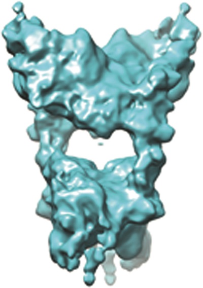
Are we blindly accepting all the interpretations that arise from our present description of X-ray diffraction?  Is it reasonable that all crystals have to be “ideally imperfect” to determine their structure?  Bragg’s law cannot avoid dynamical effects, and therefore the measured intensity is not equal to the square of the structure factor unless the crystal is assumed to be “ideally imperfect”.  If polycrystalline diffraction is formed from crystals satisfying Bragg’s law, why is the background so high compared with single crystal profiles?  Are more crystals required in polycrystalline diffraction to study complex structures with large unit cells to ensure all the peaks are captured?  If the variation of intensity around the diffraction rings from polycrystalline samples is associated with a large range of crystal sizes, why does the data from a standard reference material of similar size crystals still reveal this variation?  Are we not just modifying our sample description and instrument performance so that the current theory fits the data?  After many years of theoretical and experimental work I am convinced that I have a good explanation.

Let us look back more than a hundred years, when the two Braggs interpreted the experiments of Friedrich, Knipping and Laue.  Their interpretation was simple, clever and explained the data giving us Bragg’s law and the Bragg equation.  This equation gives the position of the diffraction peaks and any surrounding scattering is considered as a perturbation, giving information on the crystal size, strain and defects.  This description struggles to answer the questions above.

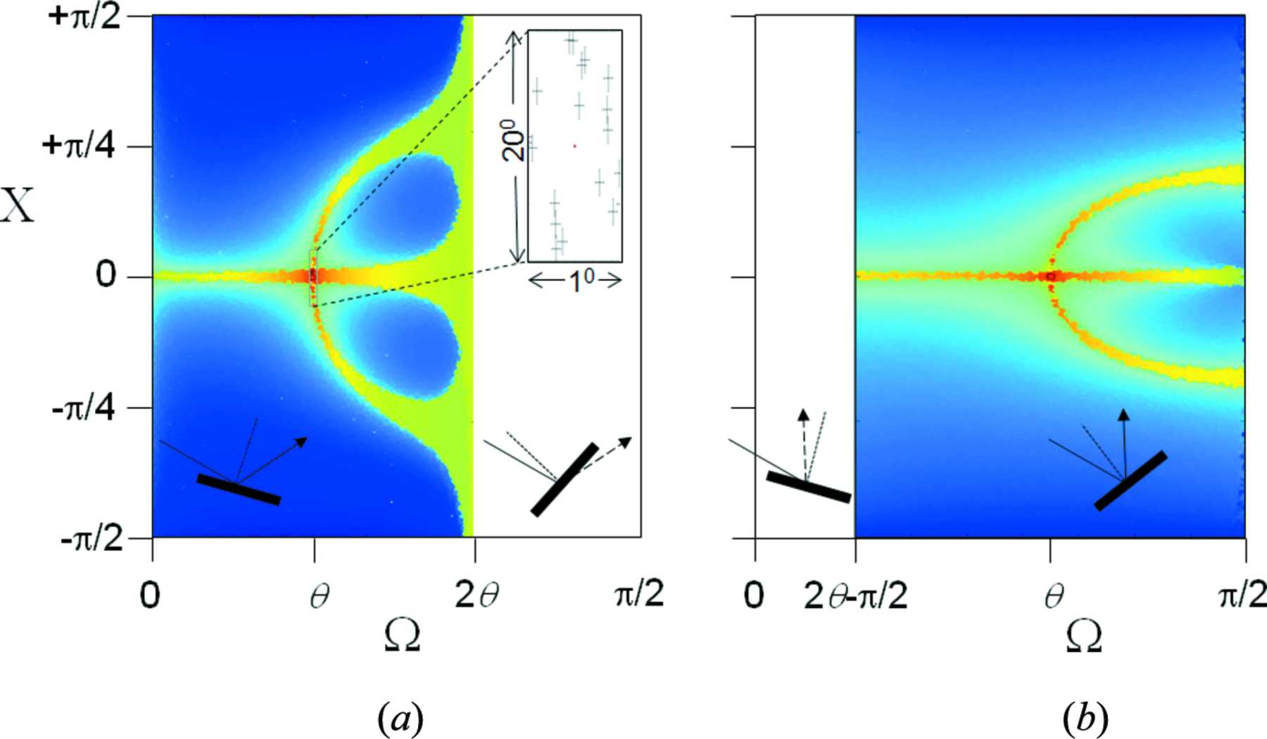
Suppose Bragg’s law is not necessary to form a diffraction peak as proposed by Fewster[1], then we can start to answer these questions.  This proposal describes how the specular (mirror) reflections from crystals planes and their periodicity give rise to two peaks, one at the mirror angle and the other at twice the Bragg angle, 2θB.  The mirror peak broadens with crystal defects and distortions, with the whole width scattering intensity towards the angle 2θB.  This broadened mirror peak contributes to the background[2].  The intensity from crystals not satisfying Bragg’s law will form a weak contribution at the angle 2θB, explaining the intensity variation in diffraction rings from polycrystalline samples.  Bragg’s law occurs where the mirror reflection and the angle 2θB peak overlap.  Therefore, if the former is broad as in an imperfect crystal Bragg’s law and therefore the dynamical effects can only exist over a small proportion of the intensity profile.

If Bragg’s law is not a requirement to create a diffraction peak, then it is possible for many peaks to be observed simultaneously.  This explains the diffraction patterns observed at X-ray free electron lasers, i.e. the appearance of several diffraction spots and their variable intensity.  Similarly, the diffraction profiles from polycrystalline materials can be explained, i.e. small numbers of crystals and the full set of peaks from a complex sample.  This description accounts for the data but indicates that a typical measurement of intensity close to a diffraction peak is inadequate, because this is only a proportion of the total intensity, and therefore cannot be directly related to the structure factor.  A study[1] on a polycrystalline silicon sample suggests that this new description gives the structural parameters within acceptable bounds whereas the conventional theory does not.

The significant step in this description is that the intensity is enhanced at the angle 2θB regardless of the crystal orientation.  This can be observed experimentally.  So why has it not been knowingly observed before?  If conventional theory is so strongly part of the crystallographer’s thinking this enhancement is easy to overlook as just some artifact.  This proposal suggests that the derived sample models could be faulty.  The magnitude of this error is difficult to assess, but with three components; the diffraction data, a theoretical description and a model of the structure, we require two of these to be correct to reproduce the third.  Suppose we assume our data is reliable, then if the theory is incomplete the structural model will be biased or unreliable.

I strongly believe that we should be questioning and discussing our current theory of X-ray diffraction, because all our structural models determined to date might be faulty or inaccurate.

[1] Fewster.  (2014).  Acta Cryst. A70, 257-282; doi:10.1107/S205327331400117X

[2] Fewster.  (2016).  Acta Cryst. A72, 50-54; doi:10.1107/S2053273315018975

View the on-demand version of our webinar with Paul Fewster the author of this work on the IUCr YouTube channel.

You can view the questions and answers covered during the webinar here.


Cryo-EM and X-ray crystallography: complementary approaches

An example of a three-dimensional structure of a macromolecule solved using cryo-electron microscopy

The invention of the electron microscope revolutionized how scientists view small structural details. The technology has undergone considerable evolution and in recent years single-particle cryo-electron microscopy (cryo-EM) has gained importance in structural biology. A topical review on cryo-EM has recently been published in Acta Crystallographica Section F (Vénien-Bryan et al., 2017, Acta Cryst. F73, 174-183). The review discusses the importance of cryo-EM and highlights recent developments. It describes how cryo-EM and other structural biology techniques, especially X-ray crystallography, now complement each other and how cryo-EM has been used in drug discovery.

The synergistic convergence of technological and computational advances now makes cryo-EM a feasible method for determining structures at near-atomic to atomic resolution (~5-2 Å). The latest generation of cryo-electron microscopes are equipped with direct electron detectors and software for the automated collection of images. In combination with the use of advanced image-analysis methods, the performance of this technique has dramatically improved. Less than a decade ago calculating a sub-10 Å resolution structure was an accomplishment but it is now common to generate structures at sub-5 Å resolution and even better. It is becoming possible to obtain high-resolution structures of biological molecules relatively quickly, in particular large ones (>500 kDa) which, in some cases, have resisted more conventional methods such as X-ray crystallography or nuclear magnetic resonance (NMR).

The potential impact of cryo-EM on drug discovery is large. Newly resolved protein structures may provide details of the precise mechanisms that are essential for cellular physiological processes. The ability to attain atomic resolution may support the development of new drugs that target these proteins, allowing medicinal chemists to understand the relationship between their molecules and targets. In addition, recent developments in cryo-EM combined with image analysis can provide unique information on connections between conformational variability and the function of macromolecular complexes.

The authors conclude that although crystallography remains the method of choice to obtain structural information from proteins for use in drug discovery, the arsenal of methods now available increases the range of possibilities, and cryo-EM is one of these methodologies, particularly for investigating changes in conformation. However, what still remains to be improved is the provision of high-quality proteins for study and so developments in purification processes are becoming fashionable once again.


Developments in the structural science of materials

C. Richard A. Catlow, Main Editor, IUCrJ

The papers published during the last year in IUCrJ in the fields of materials and computational science illustrate well the challenges posed by structural problems in the science of materials and the key role that computation can play in this and related fields in structural science. As in previous years, they demonstrate the continuing developments in techniques and instrumentation and the increasingly complex structural problems which these developments now make accessible; the role of computation in interpreting and predicting structures is equally clear.

An excellent example of technical developments facilitating new structural science is provided by the article of [Meng, Y. & Zuo, J.-M. (2016). IUCrJ, 3, 300-308], which probes three-dimensional nano-structures using a technique that employs high-resolution and low-dose scanning electron nano-diffraction (SEND) to acquire three-dimensional diffraction patterns. Their work investigates TiN – a material that is widely used in the electronics industry – and Fig. 1 illustrates how they were able to reconstruct grain structures within the material. Detailed knowledge of this microstructure is essential in understanding and optimizing the properties of the material.

Figure 1. Reconstructed grains and their orientations. Meng, Y. & Zuo, J.-M. (2016). IUCrJ, 3, 300-308

Previous editorials have emphasized the key role of diffuse scattering, which is also facilitated by technical advances. The importance of the field in materials science is well illustrated by the article of [Sawa, H. (2016). IUCrJ, 3, 298-299], which highlights the work of [Welberry, T. R. & Goossens, D. J. (2016). IUCrJ, 3, 309-318] on the interpretation of diffuse scattering from the high-temperature superconductor, HgBa2CuO4 + δ. Analysis of the diffuse scattering data reveals fascinating features involving the displacement of metal atoms around oxygen interstitial chains. This article along with several others demonstrates the need to elucidate complex structural features in disordered materials.

Analysis of diffuse scattering is also vital in the particularly exciting challenge of developing detailed models for the atomic arrangements in quasicrystals. The article of [Ishimasa, T. (2016). IUCrJ, 3, 230-231] highlights the study of [Yamada, T., Takakura, H., Euchner, H., Pay Gómez, C., Bosak, A., Fertey, P. & de Boissieu, M. (2016). IUCrJ, 3, 247-258] on the atomic structure and phason modes of the Sc–Zn icosahedral quasicrystal, which employs synchrotron-based diffraction and diffuse scattering to investigate this difficult problem.

Figure 2. A polyhedral representation of the denisovite structure. Rozhdestvenskaya, I. V., Mugnaioli, E., Schowalter, M., Schmidt, M. U., Czank, M., Depmeier, W. & Rosenauer, A. (2017). IUCrJ, 4, XXX-XXX.

The complexity of structural problem that can now be addressed is well illustrated in the paper of [Rozhdestvenskaya, I. V., Mugnaioli, E., Schowalter, M., Schmidt, M. U., Czank, M., Depmeier, W. & Rosenauer, A. (2017). IUCrJ, 4, XXX-XXX], who use a wide range of techniques including several electron crystallographic methods, XRPD and modelling to solve the structure of denisovite, a highly complex, fibrous, polytypical silicate. The structure revealed is shown in Fig. 2. The article is an elegant illustration of the capacity of, and the need for, a multi-technique approach in addressing structural problems in materials science.

A further example of complex structural science is given by the study of SnTe reported by [Sist, M., Jensen Hedegaard, E. M., Christensen, S., Bindzus, N., Fischer, K. F. F., Kasai, H., Sugimoto, K. & Brummerstedt Iversen, B. (2016). IUCrJ, 3, 377-388]. This material is increasingly investigated owing to its potential as a thermoelectric material and as a topological insulator. Their study again reveals the importance of disorder and emphasizes the need to include the effects of disorder in any theoretical investigation of the material.

Several papers illustrate both the growing power of computational methods in structural science and the role of new methodologies and algorithms in investigating structural problems [Genoni, A., Dos Santos, L. H. R., Meyer, B. & Macchi, P. (2017). IUCrJ, 4, 136-146] explore the concept of X-ray-constrained Hartree–Fock wavefunctions (XC–WF) and discuss how the procedure can be used to extract correlation effects. Their careful analysis demonstrates that the single determinant XC–WF only partially captures the effects of correlation. The paper of [Wall, M. E. (2016). IUCrJ, 3, 237-246] on quantum crystallography and the charge density of urea shows, as the authors comment, the benefits and feasibility of integrating fully periodic quantum charge-density calculations into ultra-high-resolution X-ray crystallographic model building and refinement. While the value of force-field-based methods is illustrated by the paper of [Li, X., Neumann, M. A. & van de Streek, J. (2017). IUCrJ, 4, 175-184], who evaluate different force fields in the context of their use in dynamical simulations for the prediction of chemical shifts in solid-state NMR.

The importance of the structural science of materials is, of course, illustrated by many other articles published in other journals. Of particular interest is the way in which multi-technique approaches are pinning down key structural features of catalytic materials under real operating conditions. We have previously highlighted the work of [Lezcano-Gonzalez, I., Oord, R., Rovezzi, M., Glatzel, P., Botchway, S. W., Weckhuysen, B. M. & Beale A. M. (2016) Angew. Chem. Int. Ed., 55, 5215-5219], which combines high-resolution fluorescence-detection X-ray absorption near-edge spectroscopy, X-ray diffraction and X-ray emission spectroscopy under operando conditions to provide detailed new insights into the nature of the Mo species on zeolite ZSM-5 during methane de­hydro­aromatization. Another recent example is the work of [Malta, G., Kondrat, S. A., Freakley, S. J., Davies, C. J., Lu, L., Dawson, S., Thetford, A., Gibson, E. K., Morgan, D. J., Jones, W., Wells, P. P., Johnston, P., Catlow, C. R. A., Kiely, C. J. & Hutchings, G. J. (2017). Science, 355, 1399-1403], who combined XAFS and modelling to show that in an industrially important acetyl­ene hydro­chlorination catalyst, comprising gold on a carbon support, the active sites are not, as previously thought, gold nano-clusters but single gold ions. Catalysis will unquestionably continue to pose fascinating problems for structural science.

It is hoped that this brief survey gives an impression of the range and excitement of the field of the contemporary structural science of materials and the way in which this can be unravelled by a multi-technique approach using experiment and computation. IUCrJ continues to welcome submissions in this growing field.