Dr Rosemary Wilson @rawilson80 Scientific training and outreach officer, EMBL Hamburg
 Long after the lights went on in the PETRA III Max von Laue experimental hall on the DESY campus, and the 14 beamlines became hives of activity, one corner has remained dark and seemingly forgotten. But there have always been plans for this corner of the Max von Laue Hall, and now those plans are taking shape. This hutch, situated directly behind the EMBL crystallography beamline P14, will house P14.EH2, a new experimental endstation which will cater for time-resolved crystallography experiments. In the autumn of 2016, EMBL group leader Thomas Schneider and Professor Arwen Pearson from the Centre of Ultrafast Imaging (CUI) at the University of Hamburg, were successful in securing a grant from the German federal government which in part provides funds for designing and building the endstation. Since then basic designs for the instruments have been decided, the first bits of equipment ordered and positions advertised for postdocs to help construct and run the endstation. The group hope to be able to welcome friendly users by the end of 2018. So how about some details?
Why a new endstation?
PETRA III produces some of the most brilliant and intense X-rays on the planet. The EMBL macromolecular crystallography beamline P14 is optimised to create a tight and parallel beam. With so many photons reaching the sample at the same time, scientists can start to watch the structural changes a protein goes through during a reaction, rather than a time-averaged snapshot as we are familiar with in traditional crystallography. Schneider, Pearson and their teams have already used P14 to do these types of time resolved crystallography experiments, but the set-up is not ideal in the long run.
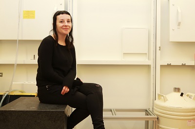
The hutch for P14.EH2 sits directly behind P14 and the new endstation will be fed by the same X-ray beam. Each time-resolved experiment will need a different instrumental set-up, optimised to the type of results the scientists want to achieve. Having a separate hutch dedicated to the complicated time-resolved set-ups means that the regular operation on P14 doesn’t need to be interrupted and the scientists can take time to plan, design and build their set-up before pressing the button to allow the X-ray beam into the hutch. “The idea is that the set-up will be very flexible and modular” explains Wellcome Postdoctoral Fellow Briony Yorke (@BrionyYorke), who is working with Pearson on the endstation. Due to the tight angle that the X-ray beam leaves the PETRA III storage ring, the other beamline hutches and endstations are narrow, with access to equipment only possible by climbing over and under beamlines and detectors. The nature of the time-resolved experimental set-up means that access has to be easy and comfortable, and this was a major consideration during the design process. (See the project webpage http://www.embl-hamburg.de/services/mx/P14_EH2 for more details on the hutch layout design and specifications.)
What methods and hardware will be available?
Time-resolved crystallography works by taking a series of snapshots of a molecule such as a protein in quick succession to build up a movie of the protein’s structural changes during a reaction (see also: Taking crystallography to the fourth dimension). In practical terms, the X-ray beam has to be interrupted to create many successive images. “The trick is to turn the X-ray beam on and off faster than the process we are interested in” explains Briony. “The current shutter on P14 is a milli-second shutter, so the only possibility to look at shorter time slices is to get the detector to count for a shorter time period by repeatedly turning it on and off, since we physically cannot open and shut the shutter fast enough.” On the new endstation, a circular metal disc with pattern of holes stamped in it called a chopper will repeatedly ‘chop’ the X-ray beam, breaking up the signal to provide the sought after shorter time slices.
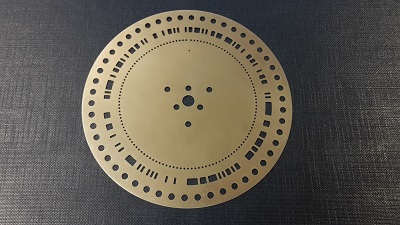
Briony’s particular interest is Hadamard time-resolved crystallography, a method she developed during her PhD, which will be an available option in the new set-up. While a chopper for standard time-resolved crystallography experiments may have a single hole in it, creating an interruption every, say second as it rotates through the beam, Hadamard choppers require a much more intricate pattern of holes. This will create many more interruptions, resulting in more data per crystal, and a more detailed ‘movie’ of the protein dynamics.
“The choppers pose quite a challenge” says Briony. “During experiments they will be spinning at very high speeds (several 1000 rpm), so they have to be cooled with water and placed under a vacuum, and shielded in a bullet-proof case something goes wrong!” Another key bit of equipment that will be found in the new hutch will be a laser for initiation of the (bio)chemical process the experiment aims to study. “The laser impulse will be delivered with fibre optics” explains Briony. “This will enable a really precise initiation of the experiments” she adds.
What science will it be used for?
“We are interested in looking at structural changes within proteins that happen during a chemical reaction, for example in an enzyme. The rate determining steps of these reactions happen on the microsecond to millisecond time-scale which P14.EH2 is designed to handle. We want to provide a regular service for people interested in exploring protein dynamics” says Briony. She will be using the time-resolved crystallography endstation, for example, to investigate how cataracts form in our eyes. “Cataracts form when proteins in our eyes start to stick together. But we don’t know what triggers this process. If we can get a picture of how and why this happens, we might be able to design medication to prevent them forming or even reverse the process” she says. P14.EH2 is likely to be interesting for a whole range of applications – from biotechnology to medicine. “For example, we could use the endstation to understand how some bacteria make crude oil, or eat plastic” explains Briony, “or gain a better understanding of how enzymes interact with medicines in our bodies. And all this evidence can be used to help build better computer simulations of molecular motions” she concludes.
What’s next?
Any day now the first instruments should arrive and the instrumentation group at EMBL can start constructing the endstation. Visit the project webpage for regular updates on their progress!
You can see papers published by Dr Thomas R. Schneider in IUCr Journals here
You can see papers published by Professor Arwen R. Pearson in IUCr Journals here