Category Archives: General

General posts not falling within the other categories of education, membership or news and opinion

IUCr2017 Hyderabad: Blog Day 2, Tuesday 22 August:The salt of the earth

Day 2, Tuesday 22 August: The salt of the earth

Dr Clare Sansom, Department of Biological Sciences,  Birkbeck College, London, UK

What links the history of crystallography, Gandhi, the Indian chemical industry and the Guinness Book of Records? Unless you keep an exceptionally strict diet, you have probably eaten some today. It is common salt, which most people will remember from high school by its chemical name: sodium chloride. This humble compound was the subject of the fascinating session that kicked off the IUCr Congress parallel programme. This programme has been designed to highlight aspects of crystallography and structural science that don’t fit into the rigid structure of plenaries, keynotes and micro-symposia that comprise the main event. ‘The salt of the earth’ fitted that bill extremely well.

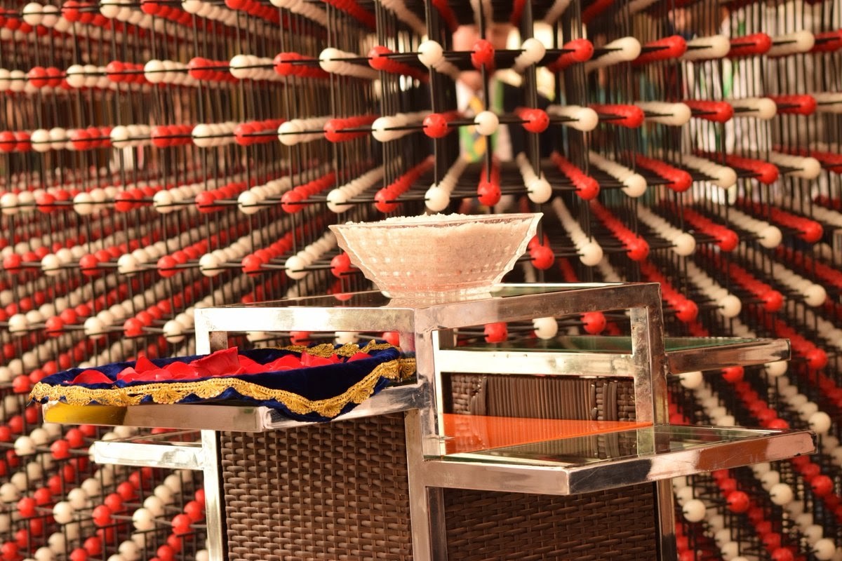
Common salt was chosen as a session topic to celebrate, of all things, a world record: the largest model crystal structure ever built. Robert Krickl, a freelance science communicator from Vienna, Austria, spent a year of his life building a 3’ x 3’ x 3’ model of a salt crystal, with its constituent sodium and chloride ions represented by red and white balls. One third of this structure will be sitting in the HICC foyer throughout the meeting. Krickl gave the first talk, and he began by explaining the inspiration behind his model. He moved into science communication following a PhD in crystallography communication and had made something of a name for himself with travelling exhibitions by the International Year of Crystallography (IYCr) in 2014. Asked then to ‘do something about it’ he was single-handedly responsible for Austria’s second place in the table of countries’ involvement in IYCr, touring the country with an exhibition and giving presentations at 182 schools. His plan to build the largest-ever model crystal structure was announced at the very end of the IYCr closing ceremony.

The choice of sodium chloride was an obvious one for Krickl; his model was built in 2015, exactly 100 years after the father-and-son team of W.H. and W.L Bragg were awarded the Nobel Prize for determining the crystal structure of this compound. A century later Krickl set about the task of acquiring the 38880 coloured balls and over 10km of sticks that he needed. The final model has repaid all his hard work, intellectually if not yet financially. If you look into it from different angles you will see an astonishing range of beautiful, symmetrical patterns. It can be disassembled so small parts can be displayed in museums or taken into schools. And the 25th IUCr congress in 2020 will be held in Prague, only about 300km from Vienna. Krickl might take his complete structure there; or, perhaps, he could challenge delegates to build a more complex model there.

Each of the four talks that followed explored a different aspect of salt: in the history of India and of crystallography, in contemporary science and in industry. Dinkar Joshi, an expert on the life of Mahatma Gandhi, described the history of salt taxes in India under the British Empire as a background to Gandhi’s famous ‘salt march’ of 1930. At the time, Indian salt was taxed to encourage Indians to buy imported salt that had been brought in as ballast for ships. Gandhi had written to the Viceroy with 11 demands, one of which concerned the salt tax; when he received no reply, he chose the salt issue for his ‘satyagraha’ (civil resistance) because of its importance to all Indians. Twenty-eight marchers, ranging in age from Gandhi himself at 61 to his teenage grandson, marched for 24 days to gather salt illegally at the coastal village of Dandi in Gujarat; the rest is history.

Mike Glazer from the University of Oxford, UK, vice-president of the IUCr explained the role that sodium chloride had played in the Braggs’ discoveries. It was not the first compound to produce a diffraction pattern (of sorts) in a beam of X-rays; that honour had gone to copper sulphate, a few years earlier. It was, however, the first to produce a diffraction pattern that was good enough for a correct structure to be determined. Interestingly, potassium chloride (KCl), which has the same structure, produced a different type of diffraction pattern because potassium and chloride ions have the same number of electrons and were therefore ‘seen’ as equivalent by the X-rays. The Braggs’ structure showed sodium chloride to be a ‘pure’ ionic compound, less than 20 years after the discovery of the electron. This was much to the chagrin of some chemists who had expected to see ‘molecules’ in the structure: but the rest, again, is history.

The final two talks brought the subject up to date. First, Artem Oganov, a solid state chemist with positions in both New York and Moscow, explained some of the properties that common salt’s ‘3D chessboard’ type cubic structure give to the many other compounds that share it. He took examples from centuries of solid state chemistry, taking the story right up to the present day and his own work. Among the structures he mentioned was a novel form of boron named gamma boron, in which small and large clusters of atoms replace the sodium and chloride ions on a cubic lattice; this is one of the hardest substances known. And finally, Amitava Das, director of an Indian government lab in Bhavnagar, described the vitally important role that common salt plays in the Indian chemical industry and thus in the country’s economy.


IUCr 2017 Hyderabad: Blog Day 1, Monday 21 August: International crystallography, collaboration and community

The opening of the 24th congress and general assembly of the International Union of  Crystallography

The opening of the 24th congress and general assembly of the International Union of Crystallography was marked by a splendid programme that stressed, time and again, the international nature of our crystallographic community. When Professor Gautam Desiraju, the organising committee chair and immediate past president of the IUCr, quoted one of the discipline’s founding fathers, W. L. Bragg, in his welcome address: “At a scientific conference nationality disappears” he could have been summing up the whole event.

Certainly, this is the most diverse IUCr congress yet, as Desiraju explained. There are representatives of 73 countries here in Hyderabad, more than the 58 national members of the International Union. India is represented by over 500 delegates and the other BRICS countries – Brazil, Russia, China and South Africa – have all sent substantial delegations. The challenges and opportunities for scientists in these very different but in many ways surprisingly similar emerging countries will be discussed later in the week. Several countries including the Cameroon and Myanmar have sent a single delegate, but the most significant example of the ‘borderless’ nature of crystallography might be the 17 delegates from Bangladesh. Three of these are students who have joined the band of enthusiastic conference volunteers on equal terms with their domestic counterparts.

The IUCr President, Marvin Hackert of the University of Texas, Austin, gave a talk running through the history of the Union and its congresses. Like independent India, the IUCr was ‘born’ in 1947, and its international congress has taken place every third year since 1948. It has grown out of recognition since those early days but its basic roles are essentially unchanged: maintaining standards, publishing books and journals (including the International Tables) and promoting international collaboration. And a number of new initiatives have been started in or after the highly successful International Year of Crystallography in 2014. These include the LAAMP project – or the Lightsources for Africa, the (Southern) Americas and Middle East Project – to provide greater access to advanced synchrotron sources for scientists working in these disadvantaged regions. India already has two such sources, with others planned.

Paul Peter Ewald, a pioneering crystallographer known to students of structural science throughout the world for his ‘Ewald sphere’, was one of those principally involved in setting up the IUCr. Since 1986, his memory has been honoured by the award of the Ewald Prize at each congress to a distinguished crystallographer. The 2017 prize was therefore the eleventh in the series; it was awarded to Sir Tom Blundell from the University of Cambridge, UK, a pioneering structural biologist and a co-founder of Astex Pharmaceuticals.

Blundell’s prize lecture was a tour de force, a whirlwind summary of half a century at the forefront of structural biology. Time and again, he came back to the importance of collaboration, both between nationalities and between academic and industrial scientists. And time and again he came back to his mentor, Nobel laureate Dorothy Hodgkin. He joined Hodgkin’s lab as a student in 1964, the year in which she won the Chemistry Nobel, and became a member of the group that solved the 3D structure of insulin. He described giving his first plenary lecture at an international conference as a 27-year-old in 1969, showing delegates at the eighth IUCr congress the newly-solved structure of that vitally important biological molecule.

During his talk, he drew out four ‘lessons’ from Dorothy Hodgkin’s example and the insulin story, illustrating them with anecdotes from throughout his career.  Firstly, he explained that good science requires a long-term vision by citing the thirty years between the first insulin crystals and the first publishable 3D structure. There are lessons here for today’s grant agencies and journal editors. His second point stressed the value of international and interdisciplinary teams and his third the importance of writing up experimental methods: Protein Crystallography, the book he co-authored with Louise Johnson in 1976, was the first authoritative textbook on this subject.

Blundell devoted most time to the fourth ‘lesson’, on the value and quality of industrial science. He has collaborated with the pharma industry since the Seventies – then perhaps rather improbably as a left-wing firebrand of an Oxford City councillor. His was one of the groups that simultaneously solved the structure of HIV’s protease, paving the way for some of the most successful drugs for AIDS. In 1999 he and two ex-colleagues, Harren Jhoti and Chris Abell, founded Astex to pioneer the technique of fragment-based drug discovery. This company is now wholly owned by Otsuka Pharmaceuticals in Japan, and eighteen years after the program was launched, its first drug, riboclicib, has just received FDA approval for breast cancer. Dorothy Hodgkin herself would recognise this time-scale. Blundell is now taking the technologies pioneered at Astex back into academia for complex targets and neglected diseases, the latter with generous funding from the Gates Foundation. Despite passing the ‘official’ retirement age he still has plenty of plans for his international, interdisciplinary team, and for what his mentor might have called ‘useful’ crystallography; I will be reporting on more of both throughout the meeting.

Dr Clare Sansom, Department of Biological Sciences,  Birkbeck College, London, UK

Quality assurance in microbeam radiation therapy

Microbeam radiation therapy (MRT) is an innovative preclinical radiotherapy procedure consisting of many micrometre-sized spatially fractionated radiation fields, obtained by collimating a beam of synchrotron radiation with a multi-slit collimator. A typical radiation field of MRT consists of an array of microbeams, each with a width of 50 µm and a centre-to-centre distance of 400 µm.

MRT differs from external beam radiation therapy (EBRT) due to the properties of synchrotron radiation, such as the small angular divergence of the photon beam, the broad spectrum of energies available and the pulsed high-intensity radiation that is produced. The low divergence of the beam ensures that the field does not spread out as it passes through the patient, thus maintaining the spatial fractionation at depth; the high-intensity radiation allows treatment time to be reduced, thus reducing smearing of the microbeam paths in the tissues due to breathing or cardiosynchronous motion.

The most significant advantage of MRT over EBRT is the different radiobiological response of cancerous and healthy tissues to the micrometre-sized MRT field. As the size of the radiation field decreases to the order of micrometres the dose tolerated by normal tissue increases dramatically, whilst maintaining tumour control. This phenomenon, called the dose-volume effect, makes MRT a promising treatment for radioresistant tumours such as osteosarcomas, or tumours located within or near sensitive structures (e.g. glioblastomas in paediatric patients).

Routine dosimetry quality assurance (QA) prior to treatment is necessary to identify any changes in beam condition from the treatment plan, and is undertaken using solid homogeneous phantoms. Solid phantoms are designed for, and routinely used in, megavoltage X-ray beam radiation therapy. These solid phantoms are not necessarily designed to be water-equivalent at low X-ray energies, and therefore may not be suitable for MRT QA.

Cameron et al. (2017). J. Synchrotron Rad. 24, 866-876 simulated dose profiles of various phantom materials and compared them with those calculated in water under the same conditions, so demonstrating quantitatively the most appropriate solid phantom to use in dosimetric MRT QA.

Based on the study, the adoption of virtual water, plastic water DT, RW3 and RM1457 solid water were recommended for MRT QA as water-equivalent solid phantom materials.


Pioneering X-ray technique to analyse ancient artefacts

A pioneering X-ray technique that can analyse artefacts of any shape or texture in a non-destructive way has been developed by an international team of scientists. The method uses X-ray diffraction (XRD) in order to determine crystallographic phase information in artefacts with very high accuracy and without causing damage to the object being scanned [Hansford et al. (2017). Acta Cryst. A73, 293-311].

Using the technique, researchers can identify pigments in paintings and on painted objects – which could potentially be applied in the future to help to clamp down on counterfeit artwork and artefacts and verify authenticity.

The research suggests that the non-invasive technique could also eliminate the frequent need to compromise between archaeological questions that can be solved and the analytical methods available to do so.

Dr Graeme Hansford, from the University of Leicester, explained, “What makes this method really unique is that the shape and texture of the sample become immaterial. I expect future studies to make significant contributions to determining the provenance of a range of archaeological objects, and this data will ultimately provide vital context information for museum collections.

“In paintings, the type of pigment used frequently yields useful insights into methods of production and the organisation of ancient industries, as well as restricting the possible date of manufacture. This could help to determine if the provenance of an artefact is as purported.”

The research was supported by the UK’s Science and Technology Facilities Council.

This news story is an excerpt taken from a press release published by the University of Leicester.


3D-printed jars in ball-milling experiments

Drawing of the Jar

Mechanochemistry is a widespread synthesis technique in all areas of chemistry. Various materials have been synthesized by this technique when the classical wet chemistry route is not satisfactory. Characterization of the reaction mixture is however much less accessible than in solutions.

Recently, in situ observations of mechanochemical reactions have been achieved by X-ray diffraction and Raman spectroscopy. Solid-state reactions can be directly tracked, revealing phase transitions and other material transformations during synthesis in a ball mill jar. This technique has become increasingly popular in different fields of mechanochemistry.

As the X-rays go through the entire jar, the diffraction patterns present a high background due to the scattering from the thick walls of the jar. Also, broad diffraction peaks are expected from the sample as a result of probing a large sample area covering the entire jar. An extra complexity arises from diffraction on the milling balls.

Tumanov et al. [(2017). J. Appl. Cryst. 50. doi:10.1107/S1600576717006744] reasoned that these issues can be resolved by modifying the geometry and material of the milling jar. But, making a jar with a complex geometry using traditional production techniques is complicated, especially at the stage of creating a prototype, when introducing changes into a design should be facile. For this reason they decided to use a 3D printer for the purpose. They show how this useful production tool can quickly make milling jars optimized for improved background, absorption and angular resolution in X-ray powder diffraction experiments; the jars are also more resistant to solvents compared with standard acrylic jars. 3D printing allows for low-cost fast production on demand.

Source files for printing the jars are available as supporting information for the paper.


Formulation of the MORPHEUS protein crystallization screens

Gorrec F, MRC Laboratory of Molecular Biology, Cambridge, UK

Technology developments, including innovative crystallization screens, are needed to obtain X-ray diffraction-quality crystals from increasingly challenging protein and other macromolecular samples. MORPHEUS crystallization screens are continuously developed to further enhance initial screening. MORPHEUS screens integrate small molecules frequently observed in the PDB to co-crystallize with proteins. These molecules are included to function as additives that act as protein stabilizers, crystal packing bridges, or any other role beneficial to protein crystallization.

Each MORPHEUS screen integrates 96 conditions, a minimal format for limiting amounts of sample. To limit the number of conditions employed, the potential ligands found in the PDB are combined into mixes. Other mixes of compounds are used, such as buffer systems and precipitant mixes that also act as convenient and effective cryoprotectants. The different types of compound mixes are combined using a fixed ratio to generate 3-D grid screens. The preparation of a screen and the optimization of conditions are amenable to automation.

A multitude of test crystallization screens are used against novel and challenging samples produced at the LMB before making choices about the formulation. The main goal is to produce exclusive crystallization hits that were not observed in other screens. MORPHEUS screens, I and II, are now used routinely in many laboratories, while MORPHEUS III is being developed. Before describing the developments related to the MORPHEUS screens, I will briefly present theoretical and pragmatic aspects of macromolecular crystallography that were taken into consideration during the early stages of MORPHEUS development. In the last section I will show how to optimize conditions following the 4-corner method.

A recording of this webinar is now available to view: https://www.youtube.com/watch?v=Gpb4SypWnQY

A complementary event is also available: https://www.youtube.com/watch?v=8cH0YNmDshY

Questions and answers raised during the webinar Formulation of the MORPHEUS protein crystallization screens

If you have any questions about the event please do not hesitate to contact Dr Jonathan Agbenyega, Business Development Manager, IUCr at ja@iucr.org


Cryo-EM and X-ray crystallography: complementary approaches

An example of a three-dimensional structure of a macromolecule solved using cryo-electron microscopy

The invention of the electron microscope revolutionized how scientists view small structural details. The technology has undergone considerable evolution and in recent years single-particle cryo-electron microscopy (cryo-EM) has gained importance in structural biology. A topical review on cryo-EM has recently been published in Acta Crystallographica Section F (Vénien-Bryan et al., 2017, Acta Cryst. F73, 174-183). The review discusses the importance of cryo-EM and highlights recent developments. It describes how cryo-EM and other structural biology techniques, especially X-ray crystallography, now complement each other and how cryo-EM has been used in drug discovery.

The synergistic convergence of technological and computational advances now makes cryo-EM a feasible method for determining structures at near-atomic to atomic resolution (~5-2 Å). The latest generation of cryo-electron microscopes are equipped with direct electron detectors and software for the automated collection of images. In combination with the use of advanced image-analysis methods, the performance of this technique has dramatically improved. Less than a decade ago calculating a sub-10 Å resolution structure was an accomplishment but it is now common to generate structures at sub-5 Å resolution and even better. It is becoming possible to obtain high-resolution structures of biological molecules relatively quickly, in particular large ones (>500 kDa) which, in some cases, have resisted more conventional methods such as X-ray crystallography or nuclear magnetic resonance (NMR).

The potential impact of cryo-EM on drug discovery is large. Newly resolved protein structures may provide details of the precise mechanisms that are essential for cellular physiological processes. The ability to attain atomic resolution may support the development of new drugs that target these proteins, allowing medicinal chemists to understand the relationship between their molecules and targets. In addition, recent developments in cryo-EM combined with image analysis can provide unique information on connections between conformational variability and the function of macromolecular complexes.

The authors conclude that although crystallography remains the method of choice to obtain structural information from proteins for use in drug discovery, the arsenal of methods now available increases the range of possibilities, and cryo-EM is one of these methodologies, particularly for investigating changes in conformation. However, what still remains to be improved is the provision of high-quality proteins for study and so developments in purification processes are becoming fashionable once again.


Developments in the structural science of materials

C. Richard A. Catlow, Main Editor, IUCrJ

The papers published during the last year in IUCrJ in the fields of materials and computational science illustrate well the challenges posed by structural problems in the science of materials and the key role that computation can play in this and related fields in structural science. As in previous years, they demonstrate the continuing developments in techniques and instrumentation and the increasingly complex structural problems which these developments now make accessible; the role of computation in interpreting and predicting structures is equally clear.

An excellent example of technical developments facilitating new structural science is provided by the article of [Meng, Y. & Zuo, J.-M. (2016). IUCrJ, 3, 300-308], which probes three-dimensional nano-structures using a technique that employs high-resolution and low-dose scanning electron nano-diffraction (SEND) to acquire three-dimensional diffraction patterns. Their work investigates TiN – a material that is widely used in the electronics industry – and Fig. 1 illustrates how they were able to reconstruct grain structures within the material. Detailed knowledge of this microstructure is essential in understanding and optimizing the properties of the material.

Figure 1. Reconstructed grains and their orientations. Meng, Y. & Zuo, J.-M. (2016). IUCrJ, 3, 300-308

Previous editorials have emphasized the key role of diffuse scattering, which is also facilitated by technical advances. The importance of the field in materials science is well illustrated by the article of [Sawa, H. (2016). IUCrJ, 3, 298-299], which highlights the work of [Welberry, T. R. & Goossens, D. J. (2016). IUCrJ, 3, 309-318] on the interpretation of diffuse scattering from the high-temperature superconductor, HgBa2CuO4 + δ. Analysis of the diffuse scattering data reveals fascinating features involving the displacement of metal atoms around oxygen interstitial chains. This article along with several others demonstrates the need to elucidate complex structural features in disordered materials.

Analysis of diffuse scattering is also vital in the particularly exciting challenge of developing detailed models for the atomic arrangements in quasicrystals. The article of [Ishimasa, T. (2016). IUCrJ, 3, 230-231] highlights the study of [Yamada, T., Takakura, H., Euchner, H., Pay Gómez, C., Bosak, A., Fertey, P. & de Boissieu, M. (2016). IUCrJ, 3, 247-258] on the atomic structure and phason modes of the Sc–Zn icosahedral quasicrystal, which employs synchrotron-based diffraction and diffuse scattering to investigate this difficult problem.

Figure 2. A polyhedral representation of the denisovite structure. Rozhdestvenskaya, I. V., Mugnaioli, E., Schowalter, M., Schmidt, M. U., Czank, M., Depmeier, W. & Rosenauer, A. (2017). IUCrJ, 4, XXX-XXX.

The complexity of structural problem that can now be addressed is well illustrated in the paper of [Rozhdestvenskaya, I. V., Mugnaioli, E., Schowalter, M., Schmidt, M. U., Czank, M., Depmeier, W. & Rosenauer, A. (2017). IUCrJ, 4, XXX-XXX], who use a wide range of techniques including several electron crystallographic methods, XRPD and modelling to solve the structure of denisovite, a highly complex, fibrous, polytypical silicate. The structure revealed is shown in Fig. 2. The article is an elegant illustration of the capacity of, and the need for, a multi-technique approach in addressing structural problems in materials science.

A further example of complex structural science is given by the study of SnTe reported by [Sist, M., Jensen Hedegaard, E. M., Christensen, S., Bindzus, N., Fischer, K. F. F., Kasai, H., Sugimoto, K. & Brummerstedt Iversen, B. (2016). IUCrJ, 3, 377-388]. This material is increasingly investigated owing to its potential as a thermoelectric material and as a topological insulator. Their study again reveals the importance of disorder and emphasizes the need to include the effects of disorder in any theoretical investigation of the material.

Several papers illustrate both the growing power of computational methods in structural science and the role of new methodologies and algorithms in investigating structural problems [Genoni, A., Dos Santos, L. H. R., Meyer, B. & Macchi, P. (2017). IUCrJ, 4, 136-146] explore the concept of X-ray-constrained Hartree–Fock wavefunctions (XC–WF) and discuss how the procedure can be used to extract correlation effects. Their careful analysis demonstrates that the single determinant XC–WF only partially captures the effects of correlation. The paper of [Wall, M. E. (2016). IUCrJ, 3, 237-246] on quantum crystallography and the charge density of urea shows, as the authors comment, the benefits and feasibility of integrating fully periodic quantum charge-density calculations into ultra-high-resolution X-ray crystallographic model building and refinement. While the value of force-field-based methods is illustrated by the paper of [Li, X., Neumann, M. A. & van de Streek, J. (2017). IUCrJ, 4, 175-184], who evaluate different force fields in the context of their use in dynamical simulations for the prediction of chemical shifts in solid-state NMR.

The importance of the structural science of materials is, of course, illustrated by many other articles published in other journals. Of particular interest is the way in which multi-technique approaches are pinning down key structural features of catalytic materials under real operating conditions. We have previously highlighted the work of [Lezcano-Gonzalez, I., Oord, R., Rovezzi, M., Glatzel, P., Botchway, S. W., Weckhuysen, B. M. & Beale A. M. (2016) Angew. Chem. Int. Ed., 55, 5215-5219], which combines high-resolution fluorescence-detection X-ray absorption near-edge spectroscopy, X-ray diffraction and X-ray emission spectroscopy under operando conditions to provide detailed new insights into the nature of the Mo species on zeolite ZSM-5 during methane de­hydro­aromatization. Another recent example is the work of [Malta, G., Kondrat, S. A., Freakley, S. J., Davies, C. J., Lu, L., Dawson, S., Thetford, A., Gibson, E. K., Morgan, D. J., Jones, W., Wells, P. P., Johnston, P., Catlow, C. R. A., Kiely, C. J. & Hutchings, G. J. (2017). Science, 355, 1399-1403], who combined XAFS and modelling to show that in an industrially important acetyl­ene hydro­chlorination catalyst, comprising gold on a carbon support, the active sites are not, as previously thought, gold nano-clusters but single gold ions. Catalysis will unquestionably continue to pose fascinating problems for structural science.

It is hoped that this brief survey gives an impression of the range and excitement of the field of the contemporary structural science of materials and the way in which this can be unravelled by a multi-technique approach using experiment and computation. IUCrJ continues to welcome submissions in this growing field.


Coordination chemistry of anions through halogen–bonding interactions

Detail of the coordination spheres of the bromide anion with Et3BuN+Br−

While an IUPAC definition of hydrogen bonding was only released in 2011 after decades of discussions in the scientific community, it did not take such a long time to come up with an analogous definition of halogen bonding, following a revival of this interaction in the literature which can be traced back to the early 1990s, Fourmigué, M. (2017). Acta Cryst. B73, 138-139

The halogen-bonding interaction is essentially described as an electrostatic interaction between a charge concentration (Lewis base) and a charge-depleted area, called an σ-hole, that a covalently bound halogen atom exhibits in the extension of this bond.

In a recent paper by Szell et al, (2017). Acta Cryst. B73, 153-162 single-crystal X-ray diffraction structures have been reported for a series of seven halogen-bonded co-crystals featuring 1,3,5-tris(iodoethynyl)-2,4,6-trifluorobenzene as the halogen-bond donor, and bromide ions (as ammonium or phosphonium salts) as the halogen-bond acceptors. Depending on the stoichiometry, the resulting frameworks can form honeycomb structures of variable geometry, but also systems with four or six halogen bonds to the bromide ion. While the counter-cations generally occupy the void spaces in the present work, the construction of halogen-bonded frameworks with potential gas storage applications is an appealing prospect which may be facilitated in the future by ligands enabling directional and multidentate interactions.


A large-solid-angle X-ray Raman scattering spectrometer

The large-solid-angle X-ray Raman scattering spectrometer at ID20. Photo credit: ESRF/McBride

S. Huotari and co-workers [J. Synchrotron. Rad. (2017), 24, 521-530] describe an end-station for X-ray Raman spectroscopy at beamline ID20 of the European Synchrotron Radiation Facility. The end-station is dedicated to the study of shallow core electronic excitations using non-resonant inelastic X-ray scattering.
X-ray Raman scattering (XRS) spectroscopy is a versatile tool for studying shallow X-ray absorption edges using hard X-rays. It has proven to be an invaluable technique for the study of electronic excitations in a variety of sample systems such as crystals, liquids and gases. Over the past decades, XRS has been applied to solve geoscientific questions by studying shallow core edges under extreme pressure and temperature conditions, follow chemical reactions in situ, and study liquid samples under well defined thermodynamic conditions.
A drawback of XRS is the orders-of-magnitude weaker scattering cross section in comparison with the probability for photoelectric absorption. This can be compensated for by using light sources with a very high brilliance and efficient signal collection; this has been the guiding motive for the design of the spectrometer presented in this paper.
The new end-station provides an unprecedented instrument for X-ray Raman scattering, and will open the door to renewed studies of low-energy X-ray absorption spectra in materials under in situ conditions, such as operando batteries and fuel cells, in situ catalytic reactions, and extreme pressure and temperature conditions.